
The Norwood scale classifies male pattern hair loss into seven main stages, from little or no visible recession to extensive loss across the top of the scalp. Also called the Hamilton-Norwood scale, it describes the hairline, temples and crown. It helps you discuss a pattern with a clinician, but it cannot establish the cause of hair loss, predict your final stage or calculate your graft requirement by itself. Original classification · Classification review
If your photos seem to sit between two stages, you do not need to force a number. Start with clear pictures, changes over time and a scalp assessment. Choosing treatment is a separate decision.
Norwood scale in 60 seconds
- There are seven main stages, I to VII. Stage III Vertex and Type A describe additional patterns.
- Norwood 2 can resemble a mature hairline. Continued recession or miniaturization needs assessment.
- Norwood 3 is the conventional threshold for established balding in the chart. Earlier changes can still deserve medical attention.
- Your crown matters. A frontal selfie can miss a vertex pattern.
- Your stage does not set your future. There is no reliable stage-per-year timetable for an individual.
- Your donor area needs its own assessment. A stage number cannot determine transplant eligibility, graft count or price.
Discuss the changes you have noticed and the assessment that could clarify them.
What is the Hamilton-Norwood scale?
The Hamilton-Norwood scale is a visual classification of the distribution and extent of male pattern hair loss, or androgenetic alopecia. James Hamilton described an earlier system in 1951. O’Tar Norwood refined the classification in 1975. Arabic numbers, such as Norwood 3, and Roman numerals, such as Norwood III, refer to the same main stage. Norwood’s paper
Male pattern loss involves genetic and hormonal sensitivity in susceptible follicles. It can produce miniaturization before the pattern becomes obvious in a selfie. Our DHT and hair loss explanation covers that mechanism. BAD patient guidance
A higher stage generally describes more extensive patterned loss. The numbers are categories, not measured percentages of missing hair. They also do not measure daily shedding, follicle health or how distressed someone feels about their appearance.
What do the seven Norwood stages look like?
Use the hairline and the crown together. The bridge is the band of hair across the middle of the scalp that can separate frontal and crown loss. These descriptions follow the conventional chart; a clinician may document a pattern that does not fit neatly. Institute of Trichologists guide
| Stage | Main visible pattern | Useful distinction |
|---|---|---|
| Norwood 1 / I | Little or no significant temple recession | A low stage does not measure density throughout the scalp. |
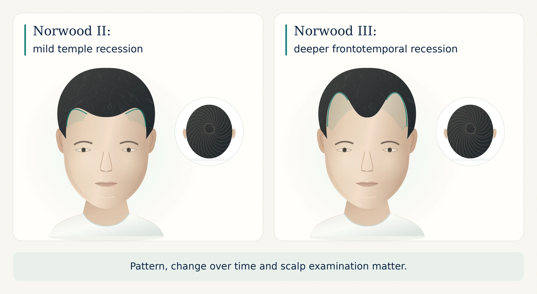
| Norwood 2 / II | Mild recession at the temples | May resemble a mature hairline; check change over time. |
| Norwood 3 / III | Deeper frontotemporal recession | Classic stage III does not require a crown bald spot. |
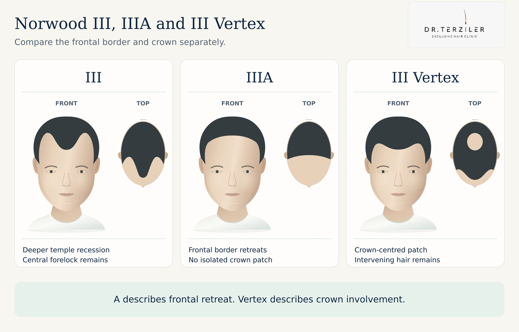
| Norwood 3 Vertex / III V | More obvious thinning at the crown, with frontal recession no more advanced than III | Vertex is a pattern variant, not an eighth main stage. |
| Norwood 4 / IV | More extensive frontal and crown loss | A substantial band of hair still separates the two areas. |
| Norwood 5 / V | Larger frontal and crown areas | The separating band becomes narrower and sparser. |
| Norwood 6 / VI | Frontal and crown loss join | The separating bridge has disappeared. |
| Norwood 7 / VII | Most extensive loss over the top | A relatively narrow horseshoe of hair remains at the sides and back. |
What does Norwood 1 mean?
Norwood 1 describes little or no significant recession in the conventional pattern. Hairlines naturally vary, so it does not require a perfectly straight juvenile hairline. Someone can have a stage I outline while noticing reduced density elsewhere. A full-looking frontal border alone cannot exclude another form of thinning.
Is Norwood 2 a mature hairline or early balding?
Norwood 2 describes mild recession at the temples. That shape can be consistent with a mature adult hairline, but the number does not prove that the hairline has stabilized. A change in shape, reduced density or finer hairs may justify assessment even when the recession looks modest.
Compare photographs taken under similar conditions. A clinician can examine miniaturization, which means follicles producing progressively finer hairs. Family history helps, but it does not settle the diagnosis. How male hair loss is assessed
For a closer comparison, read our mature versus receding hairline guide.
What does a Norwood 3 hairline look like?
Norwood 3 has deeper recession at the frontal corners than stage II. The remaining central hair may create an M-shaped outline. This is the conventional minimum stage described as established balding in Norwood’s classification. It does not mean that earlier androgenetic changes are medically irrelevant.
A prominent widow’s peak is not enough to diagnose stage III. The extent of recession and the surrounding hair matter more than whether the outline resembles a particular letter.
What is Norwood 3 Vertex?
Norwood 3 Vertex, often written III V or 3V, describes more apparent crown loss with frontal recession no more advanced than stage III. The vertex is the upper back or crown of the scalp. Classic stage III and III Vertex should be recorded separately because they describe different distributions. Classification reference
What changes at Norwood 4 and 5?
At Norwood 4, frontal recession and crown loss are more extensive, while a band of hair separates them. At Norwood 5, the areas enlarge and that band becomes thinner and narrower. Comparing the bridge helps distinguish these stages, although photographs and hair length can make the boundary difficult to judge.
What distinguishes Norwood 6 from 7?
At Norwood 6, frontal and crown loss have joined across the top. At Norwood 7, loss is more extensive and the remaining side-and-back fringe is narrower. Neither stage tells you whether that remaining hair is a safe surgical donor. Donor density, miniaturization, previous harvesting and scalp condition require separate examination. Donor assessment
How can you tell Norwood 2 from Norwood 3?
The main difference is the depth and extent of frontotemporal recession. Norwood 2 shows a mild retreat at the corners. Norwood 3 shows more established recession. There is no universal finger-width or forehead-centimeter test that accurately makes the distinction for every face.
 Open full-size image
| Question | What to compare |
|---|---|
| Is the hairline changing? | Similar-angle photographs from different dates. |
| Are the corners deeper? | Both temple views, without stretching the skin or changing head tilt. |
| Is density changing behind the border? | Scalp examination and, when appropriate, trichoscopy. |
| Is the crown also thinning? | A separate top and crown view. |
| What if the pictures are inconclusive? | Record uncertainty and ask for an assessment. |
Unsure whether you are a 2 or a 3? Bring comparable pictures and ask what the examination changes about your next step.
Bring comparable photographs and ask what a scalp examination changes about your next step.
What are Norwood Type A, 3A and 3 Vertex?
Type A describes a more uniform retreat of the frontal hairline. In the classic Type A variants, the front recedes backward without the usual separate early crown bald spot or persisting central forelock. The variants are generally recorded as IIA, IIIA, IVA and VA. They are not extra numbered stages after VII.
Norwood 3A and Norwood 3 Vertex describe different patterns. IIIA concerns the frontal retreat. III Vertex highlights crown involvement. A front-only photograph may be inadequate to tell them apart. Review of the variants
 Open full-size image
You may also see “Norwood 2 Vertex” online. Describe the actual crown change to a clinician rather than assuming that every internet label is a formal category.
How can you check your Norwood stage at home?
A home check can help you describe what you see. It cannot confirm androgenetic alopecia or replace a scalp examination.
-
Take a straight-on hairline photograph. Keep the head position natural and the whole frontal border visible.
-
Photograph both temples. Use matching angles and avoid pulling the hairline unevenly.
-
Take a top-down view. Include the middle of the scalp rather than the hairline alone.
-
Add a crown view. The crown whorl may need a different angle from a front-facing top photo.
-
Add the back and sides for a consultation. These views help the clinician decide whether a donor assessment is needed; photos alone cannot establish harvesting safety.
-
Match the conditions next time. Keep lighting, camera distance, hairstyle, hair length and wetness as consistent as practical. Record the date and any treatment changes.
Researchers have published standardized photography protocols because inconsistent images make hair-loss assessment harder. This home checklist is a practical adaptation, not a validated diagnostic test. Clinical photography guide
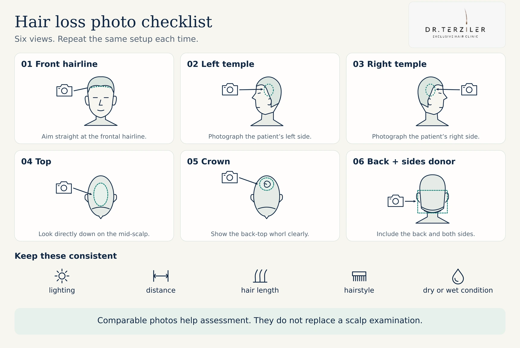 Open full-size image
Why do wet hair and bathroom lighting change the picture?
Wet or oily hair groups into strands. Directional light and camera exposure change how much scalp you see. Hair fibers, styling products, a new haircut or a different part can also change the apparent coverage. Those differences can make a comparison misleading without proving that your stage changed.
Use comparable photographs to discuss a change. Arrange an earlier assessment if you have sudden shedding, patches or scalp symptoms rather than waiting to build a perfect photo diary.
How accurate is a Norwood scale test or AI calculator?
A Norwood test is usually a picture-matching exercise. Its usefulness depends on the views, image quality, categories and person or system interpreting the result. A score such as “Norwood 2.37” suggests more precision than the conventional classification supports.
A published study of 105 men aged 30 to 70 compared self-ratings with ratings by trained observers. Men’s exact agreement with the observers was 48% to 55%. The two trained observers agreed exactly in 85% of cases. This was a specific Australian study, not an accuracy score for every modern quiz or app. Study of self-reported balding patterns
Professional interpretation also has limits. A separate study in which 23 dermatologists and dermatology residents rated 43 photographs found imperfect agreement and poor repeatability in a second assessment exercise. Treat a borderline category as a reason for better assessment, not as a precise measurement. Hamilton-Norwood reliability study
An AI hair-loss tool should explain which views it needs, who supplied its reference labels, how it was tested and what happens when it cannot classify an image. Research on an AI model does not validate every consumer app using the word AI. An app also needs a clear policy for storing or deleting identifiable scalp photographs.
Want a useful answer beyond a score? Ask whether the pattern is clear, whether another diagnosis needs consideration and what the next assessment should include.
Ask what is clear, what is uncertain and what needs an examination.
What if you have diffuse thinning, a normal hairline or a visible crown whorl?
The Norwood outline can underrepresent hair loss spread across the top of the scalp. A person may retain a recognizable hairline while density falls behind it. Diffuse thinning also has several possible causes, including temporary shedding and other disorders. A clinician may use additional descriptions, photographs and scalp measurements instead of forcing the appearance into a stage.
A crown whorl naturally exposes some scalp where hairs change direction. One bright photograph cannot reliably distinguish that appearance from crown thinning. Change over time and the examination are more useful than a single circle of visible skin.
If thinning extends into the back and sides, the donor area needs particular attention. Diffuse unpatterned alopecia, often called DUPA, may undermine the assumption of a stable donor zone. It must be distinguished from other diffuse conditions. 2026 clinical review
Our hair loss guide explains the wider diagnostic pathways. A digital scalp assessment may add information about hair caliber, density and miniaturization where clinically appropriate.
Does the Norwood scale apply to women?
The Norwood scale was developed around male-pattern distributions. Female pattern loss more often involves reduced density across the central scalp, and clinicians commonly use Ludwig or Sinclair descriptions. Some women have frontal or temporal recession, so the visible pattern and cause still need individual assessment. Sex alone does not make every presentation fit one chart. Classification systems
The basic and specific, or BASP, system is another approach that can describe different hairline shapes and density patterns. A comparative study found better agreement with BASP than with Hamilton-Norwood in its assessment setting. No chart replaces diagnosis. Classification reliability comparison
Will Norwood 2 or 3 always progress to Norwood 7?
No. Your current stage cannot establish your eventual stage or the speed of future loss. Pattern hair loss may progress over years or decades, and individual courses differ. Age at onset, clinical history, treatment and changes on examination help a clinician discuss risk, but they do not supply an exact countdown. British Association of Dermatologists guidance
There is also no single “normal Norwood stage” for every age. A stage at 20 and the same stage at 50 carry different histories, but neither age alone determines whether medication or surgery is appropriate. A relative’s photograph cannot predict your future hairline precisely.
If an online chart gives an average age for each stage, check which population it studied. Population prevalence and an individual prediction answer different questions.
Can treatment reverse a Norwood stage?
Treatment may improve density, reduce ongoing loss or alter the visible pattern in some people. It cannot guarantee a return to Norwood 1. A person can also benefit from treatment while remaining in the same broad stage because these categories do not capture every density change.
Medical options include minoxidil and finasteride for appropriately selected patients. They have different mechanisms, precautions and side effects. Preserving existing hair and restoring a missing border are different treatment goals. AAD treatment guidance
| Finding | What an assessment may clarify |
|---|---|
| Mild recession with uncertain change | Whether observation, comparable photographs or further examination is enough. |
| Active miniaturization or progressive pattern loss | Whether medical treatment is suitable and how response will be measured. |
| Patchy loss, heavy sudden shedding or scalp inflammation | Whether another diagnosis needs investigation before cosmetic treatment. |
| Established loss with a suitable donor area | Whether surgery can achieve the agreed coverage without exhausting donor resources. |
| Extensive loss with limited donor supply | Which areas can realistically be prioritized, or whether surgery should be avoided. |
Does minoxidil or finasteride work for a receding hairline?
Response varies by person, site and product. Some US topical minoxidil labels specify vertex use and state that the product is not intended for frontal baldness or a receding hairline. That labeling should not be turned into a claim that every hairline will respond, or that all clinician-directed uses are identical. Example US product label
Discuss finasteride’s potential benefits and adverse effects with a prescriber. US compounded topical finasteride is not FDA-approved, and topical application does not eliminate the possibility of systemic effects. FDA safety information
What about PRP, laser treatment or a hair transplant?
These options serve different purposes and have different evidence. They should not be assigned automatically to a Norwood number. Our PRP hair treatment guide explains the research and its limitations. A transplant moves existing follicles and requires a plan for future native-hair loss. NHS transplant guidance
How many grafts do you need at each Norwood stage?
There is no dependable one-to-one conversion from a Norwood stage to a graft count. The stage describes a pattern; the surgical plan also requires measurements and an assessment of the donor supply. ISHRS illustrates why people in the same classification may need different plans. ISHRS planning explanation
For a simple arithmetic example, imagine two restoration areas discussed at an assessment:
| Worked example | Area | Illustrative added graft density | Arithmetic |
|---|---|---|---|
| Smaller planned area | 80 cm² | 30 grafts/cm² | 2,400 grafts |
| Larger planned area | 120 cm² | 30 grafts/cm² | 3,600 grafts |
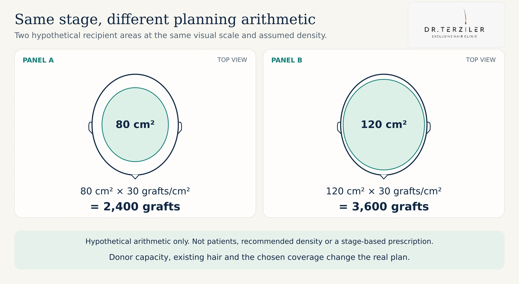 Open full-size image
These are hypothetical planning examples, not patient records, recommended densities or stage-based graft ranges. They illustrate why the same nominal density needs 50% more grafts when the planned area is 50% larger. Existing hair, hair caliber, graft composition, safe placement and donor capacity change the real plan. Hair counts and graft counts are not interchangeable.
Ask for the measured areas, the planned distribution and the donor assessment behind a number. Our graft estimation guide explains those inputs.
Before you compare graft quotes, compare the plan. Ask what can be covered now, what density is realistic and what donor resources remain for later.
Measured areas, donor findings and realistic coverage belong in the same discussion.
Is a hair transplant possible at Norwood 6 or 7?
It may be possible for selected people, but a high stage neither guarantees nor automatically rules out surgery. Extensive recipient loss increases the importance of donor capacity and realistic priorities. A plan may emphasize the hairline and frontal scalp, accept lighter coverage elsewhere or involve staged treatment. Body hair is an option only in selected cases, with its own limitations.
Sapphire FUE, DHI or a robotic system cannot create an unlimited donor supply. Ask who will assess the donor, plan the hairline and take responsibility for the procedure. Learn about doctor-led hair transplant planning in Turkey and compare written package costs and inclusions after an assessment. A price or graft number on a stage chart is not your surgical quote.
Real patients: different patterns, individual plans
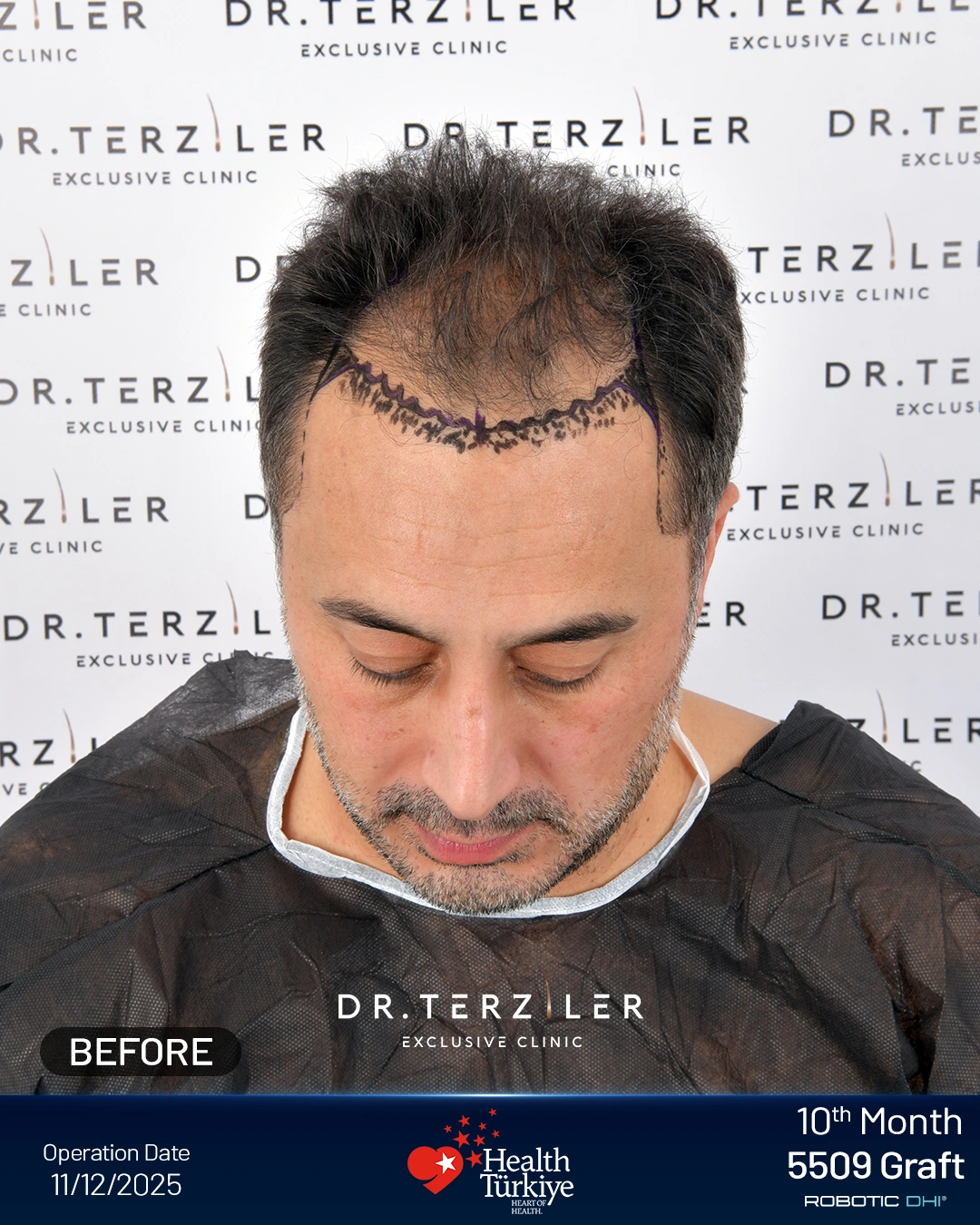
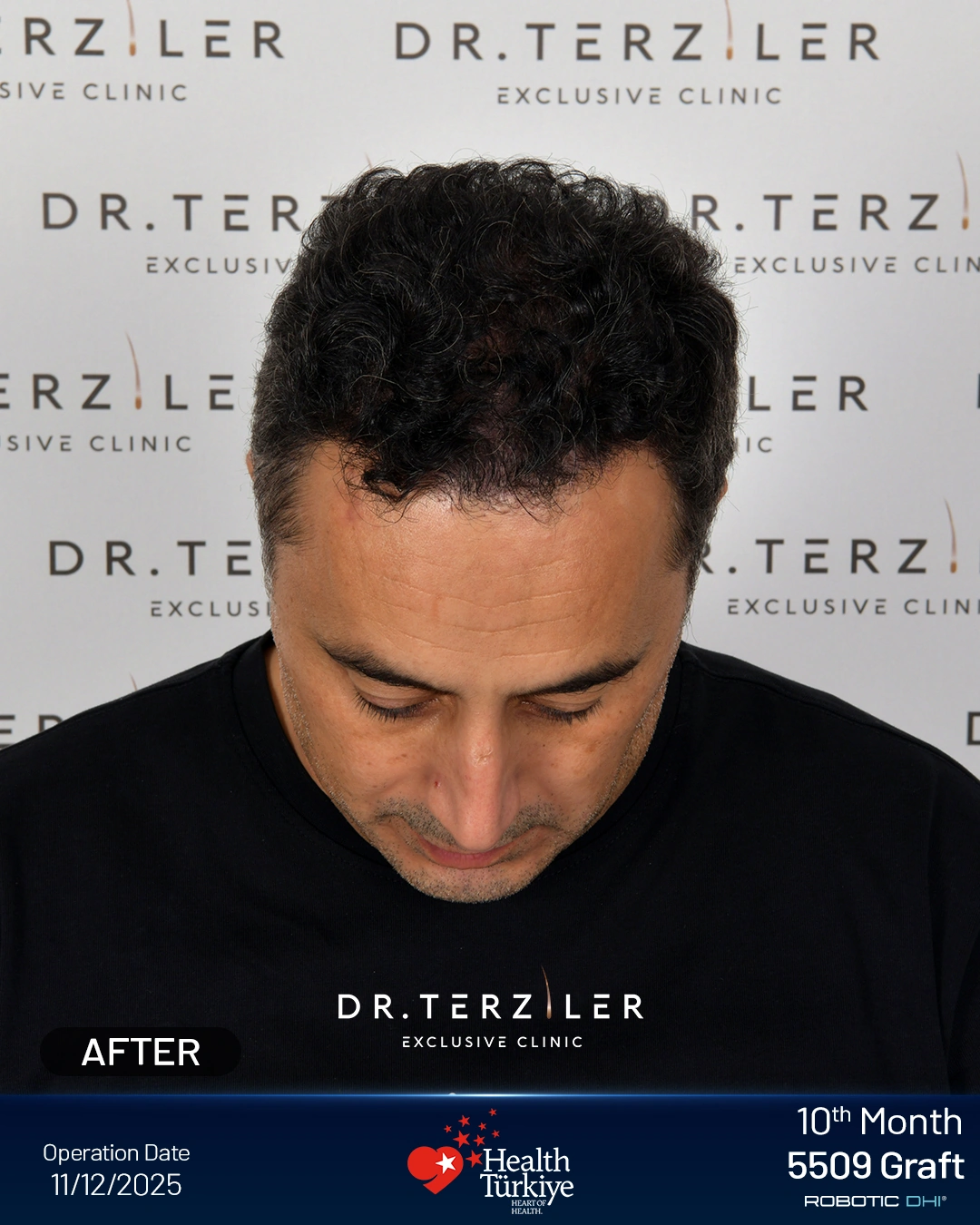
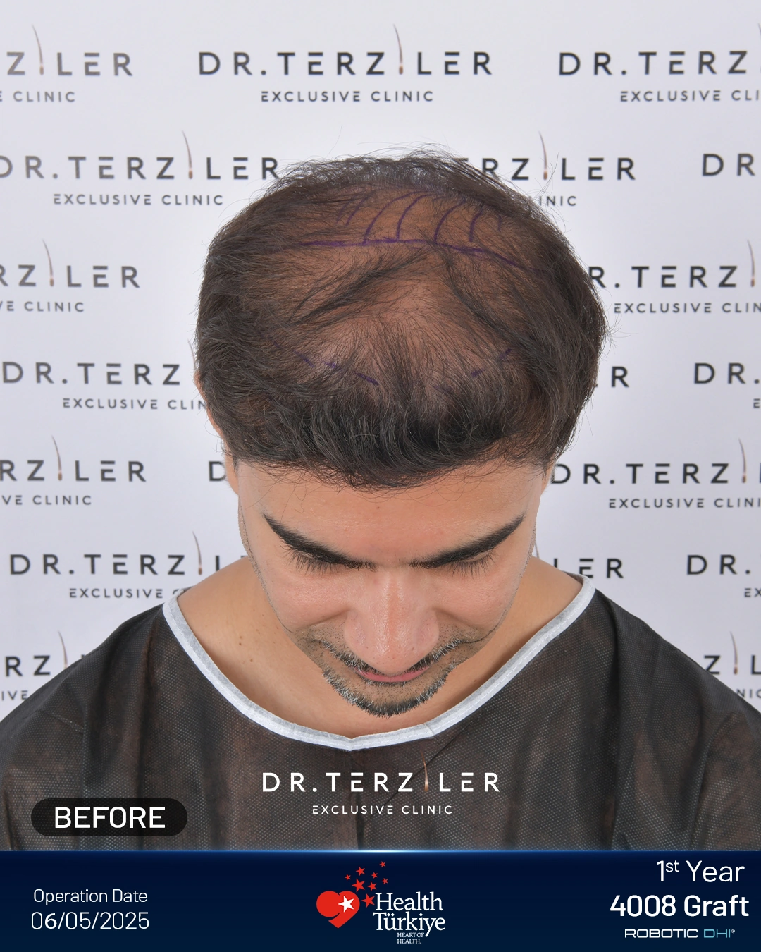
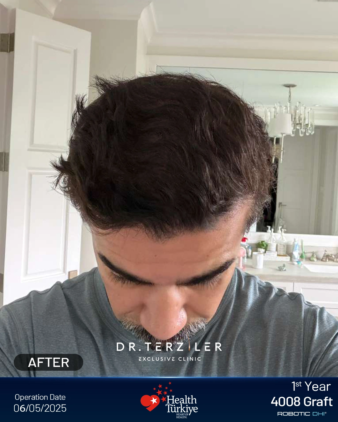
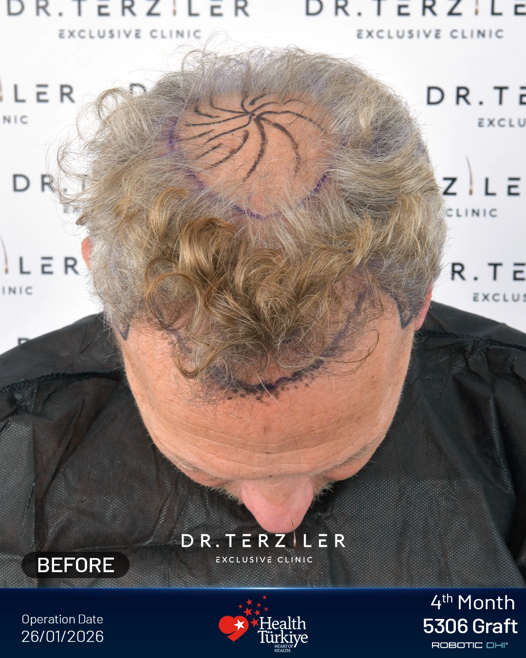
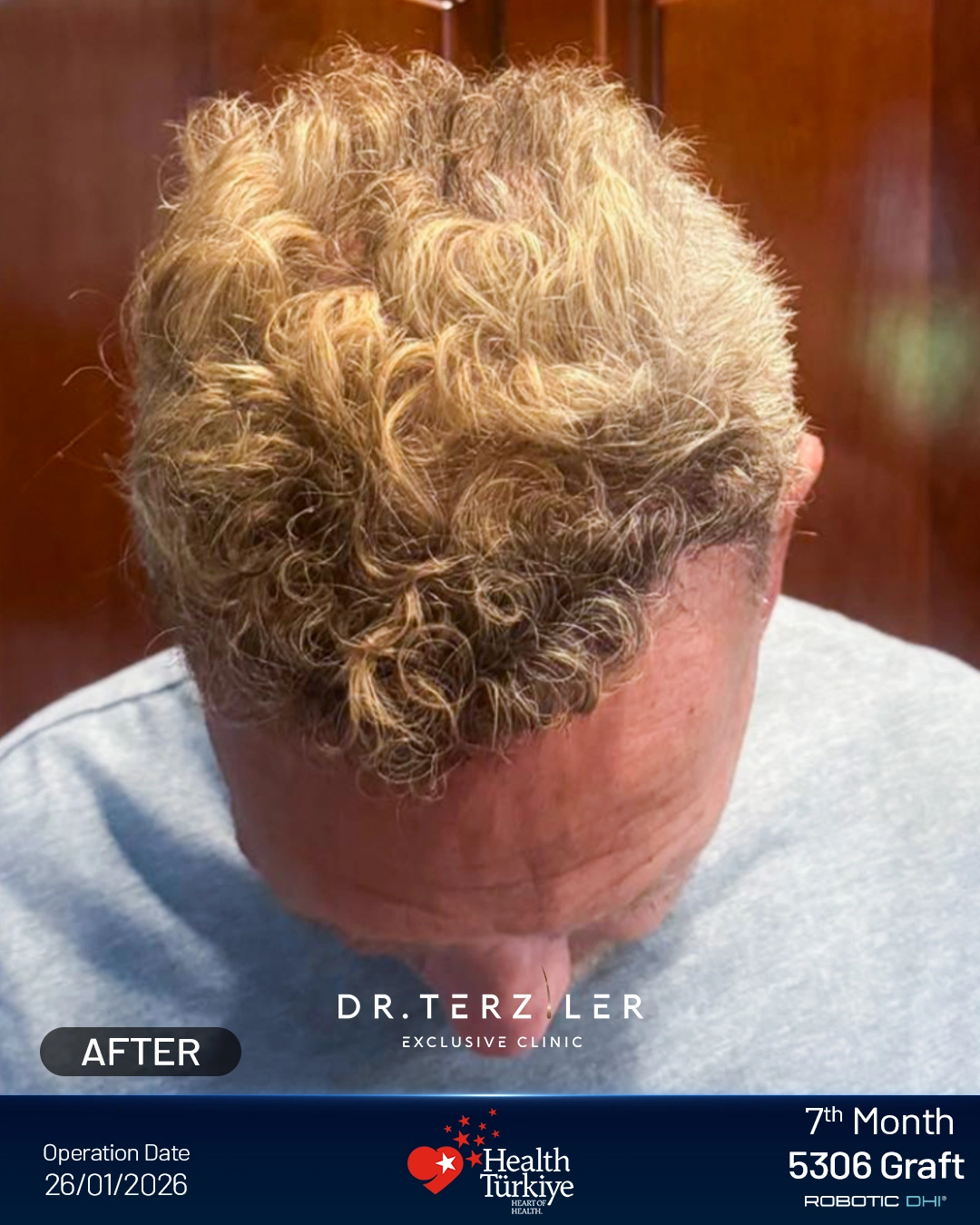
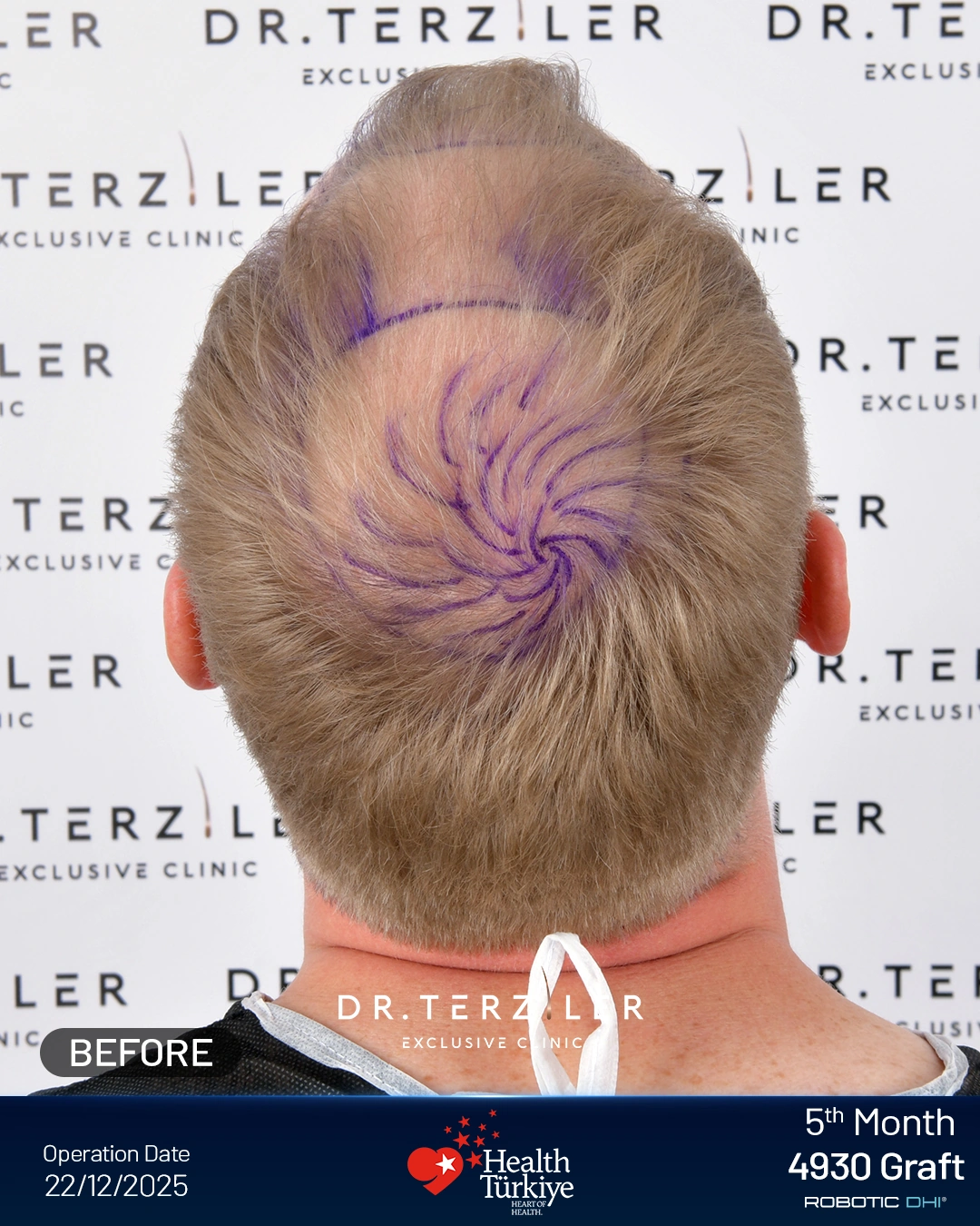
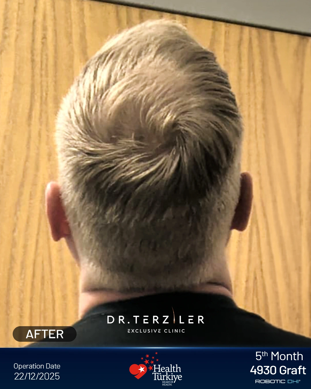
These six clinic-provided examples show visible hair-loss patterns and follow-up appearances. Case numbers identify the photographs, not Norwood stages. Graft counts and follow-up times are the clinic’s labels; the photographs do not measure graft survival or predict your result.
6 examples shown
Frontal hairline
4,159 grafts 7-month follow-up
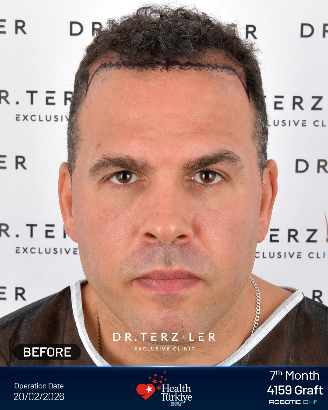
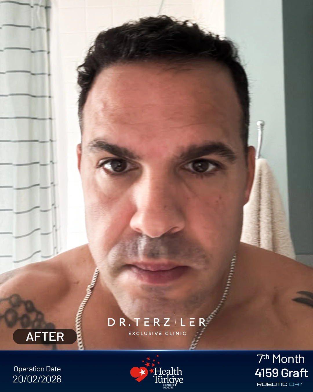
Frontal thinning
5,509 grafts 10-month follow-up
Diffuse top thinning
4,008 grafts 1-year follow-up
Front and crown thinning
5,306 grafts 7-month follow-up
Crown thinning
4,930 grafts 5-month follow-up
Advanced frontal recession
4,014 grafts 1-year follow-up
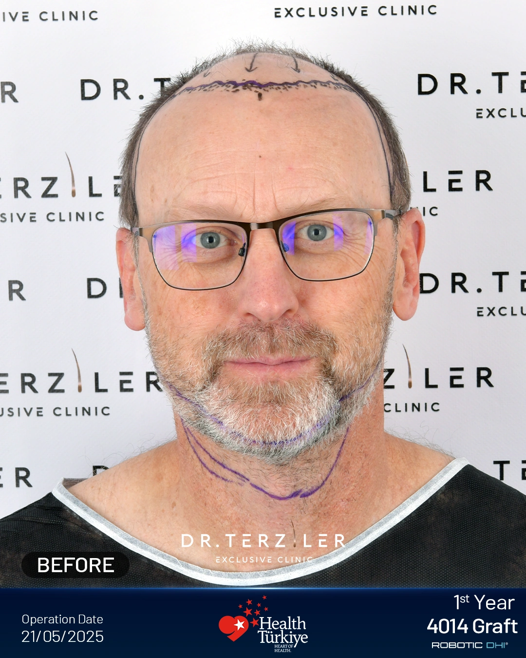
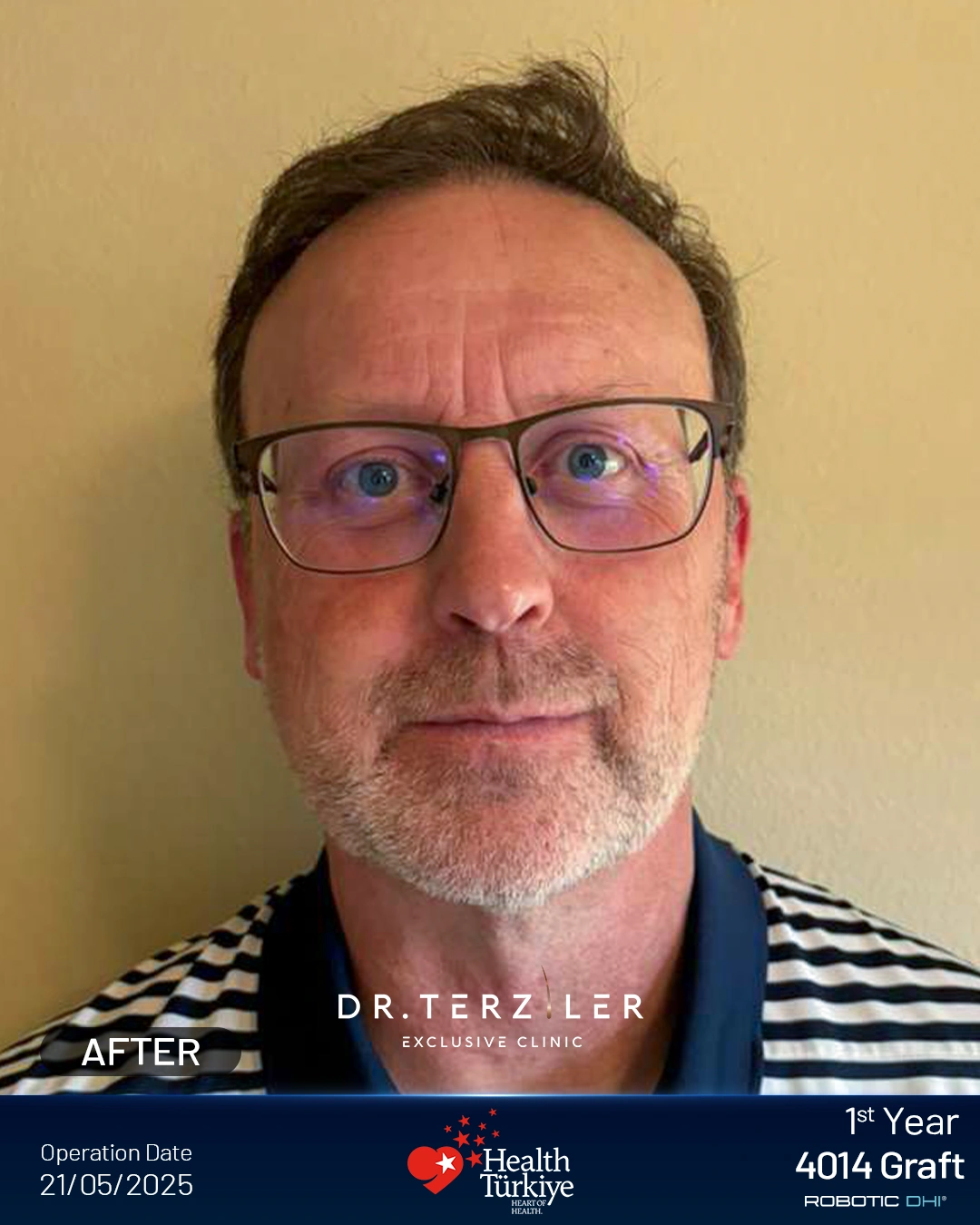
Five- and seven-month photographs are follow-up examples, not automatically final outcomes. Exact baseline Norwood stages have not been established from these photographs. A graft number from one case is not a stage-based prescription.
Ask what your donor assessment, existing hair and coverage priorities change about your options.
What did our first-consultation records show about Norwood stages?
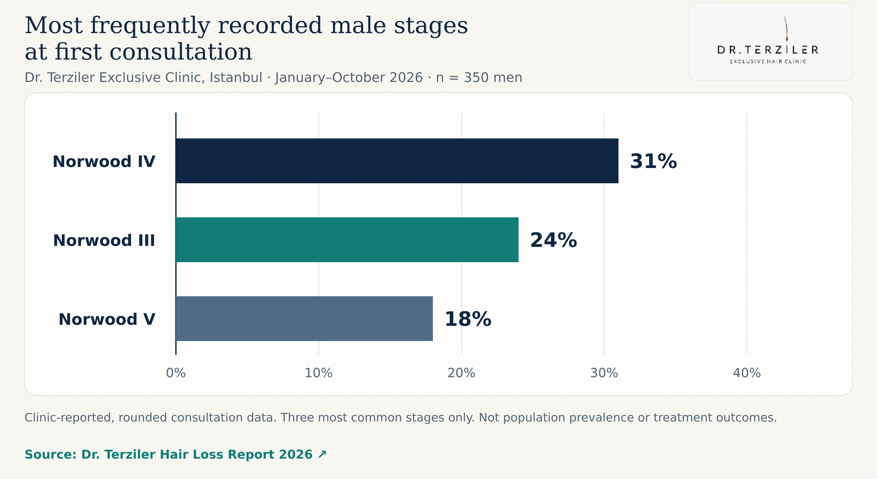
In Dr. Terziler Exclusive Clinic’s reported review of 500 first-consultation patients in Istanbul from January to October 2026, the male subgroup included 350 men. The most frequently recorded stages were:
| Recorded male stage | Share of the 350 men |
|---|---|
| Norwood 4 | 31% |
| Norwood 3 | 24% |
| Norwood 5 | 18% |
 Open full-size image
These are clinic-reported, rounded figures from people seeking a consultation. They describe the three most frequently recorded stages, not the complete distribution or the prevalence of baldness in the US, UK or Turkey. They also do not show how many of those men required surgery.
Across the full 500-person cohort, which included 350 men and 150 women, 19% were advised against a transplant at the first visit. Reported reasons included active shedding, a weak donor area and rapidly progressing loss in younger patients. This does not mean that every patient was permanently unsuitable.
Source and methodology: Dr. Terziler Hair Loss Report 2026. These consultation observations are not a patient-satisfaction survey or a treatment-outcome study.
What should a Norwood consultation actually answer?
A useful consultation should explain the pattern, the likely diagnosis and the decision it supports. At Dr. Terziler Exclusive Clinic, you can request an assessment and discuss the plan with the medical team. Ask which named physician will be responsible for your case and what further examination is needed.
Bring the onset date, the speed of change, comparable photographs, current medications and previous treatment history. Ask:
You can view the profiles of Dr. Servet Terziler and Dr. Kürşat Yalvaç before arranging an appointment.
When should you stop comparing charts and see a doctor?
Arrange medical assessment for sudden or unusually heavy shedding, distinct patches, scalp pain, burning, marked redness, pustules or signs of scarring. Loss involving eyebrows or body hair also needs a broader assessment. Do not delay that assessment because an online stage looks low. AAD diagnosis and treatment guidance
If repeated checking is affecting sleep, concentration or daily life, discuss that distress too. You deserve help with how the concern feels as well as how the scalp looks.
Norwood scale FAQ
21 questions shown
Is Norwood 2 always balding?
Is Norwood 3 too late to save my hair?
Can I stay Norwood 2 forever?
What are Norwood 0, 1.5, 2.5 and 8?
Can I be Norwood 1 and still have thinning hair?
Does Norwood 3 Vertex mean the same as 3A?
Is a visible crown always a bald spot?
Is there a normal Norwood stage for my age?
How long does Norwood 2 take to become Norwood 3?
Can minoxidil move me down the Norwood scale?
Can you get a hair transplant at Norwood 2?
Is Norwood 7 too bald for a transplant?
How many grafts does Norwood 4 need?
Can a photo or AI app diagnose my hair loss?
Can women use the Norwood scale?
Will shaving my head change my Norwood stage?
Why does one temple look worse than the other?
Can treatment work even if my Norwood number stays the same?
Should I check my stage every day?
What do “norwooding” and “Norwood reaper” mean?
Does Norwood 4 mean I have lost four-sevenths of my hair?
Get a plan that explains your pattern
Bring the photos you have and the questions you want answered. We can help you arrange an assessment and discuss whether monitoring, medical treatment or a donor-based surgical plan is the appropriate next step.
Ask whether monitoring, treatment or a donor-based surgical plan is appropriate.
The useful result is a decision you understand, including when the right decision is to wait.
Sources and further reading
- Norwood OT. Male pattern baldness: classification and incidence. 1975. Original paper record.
- Gupta M, Mysore V. Classifications of Patterned Hair Loss: A Review. 2016. Open-access review.
- The Institute of Trichologists. The Norwood Scale best-practice guide. PDF.
- Taylor R et al. Validity of self reported male balding patterns in epidemiological studies. 2004. Study record.
- Guarrera M et al. Reliability of Hamilton-Norwood Classification. 2009. Full text.
- Reliability of the pattern hair loss classifications: a comparison of BASP and Norwood-Hamilton. Study record.
- Takwale A et al. A practical guide to the standardization of hair loss photography for clinicians. 2025 issue, published online 2024. Study record.
- Classification of Male-pattern Hair Loss. 2017. Open-access article.
- American Academy of Dermatology. What is male pattern hair loss, and can it be treated? Patient guidance.
- American Academy of Dermatology. Hair loss: diagnosis and treatment. Patient guidance.
- British Association of Dermatologists. Male pattern hair loss patient information. Guidance and downloadable leaflet.
- ISHRS. The donor site: evaluation and planning. Professional society explanation.
- ISHRS. Scalp Scan Video: why the same Norwood stage can need different plans. Planning explanation.
- NHS. Hair transplant. Suitability and limitations.
- FDA. Risks associated with compounded topical finasteride. Safety communication.
- DailyMed. Example US minoxidil label. Product-specific labeling.
- Revisiting Diffuse Unpatterned Alopecia: Reappraisal of a Controversial Diagnosis. 2026. Clinical review record.
- Dr. Terziler Exclusive Clinic. Hair Loss Report 2026. Clinic-reported first-consultation findings and limitations.
- Internet terminology background only: Norwood scale and Norwood reaper meme documentation. This is not a medical evidence source.